Review Article
DOI: http://dx.doi.org/10.18203/issn.2454-2156.IntJSciRep20194253
Serum lipids and lipoproteins: a brief review of the composition, transport and physiological functions
Department of General Surgery, MM University, Mullana, Ambala, Haryana, India
Received: 26 June 2019
Revised: 03 September 2019
Accepted: 05 September 2019
*Correspondence:
Dr. Sharadendu Bali,
E-mail: drsharadbali@yahoo.com
Copyright: © the author(s), publisher and licensee Medip Academy. This is an open-access article distributed under the terms of the Creative Commons Attribution Non-Commercial License, which permits unrestricted non-commercial use, distribution, and reproduction in any medium, provided the original work is properly cited.
ABSTRACT
The dietary fats are composed primarily of triacylglycerols and some amount of phospholipids and cholesterol. Being hydrophobic in nature, these are insoluble in water, and hence cannot be transported in the blood plasma per se; to enable these lipids to be transported by the blood stream to various peripheral tissues, nature has devised the technique of making these soluble by binding them to proteins. These proteins involved in lipid transport are known as apolipoproteins, and the protein-lipid particle is known as lipoprotein. Thus, lipoproteins can be considered to be the primary transport mechanism to carry lipids from the alimentary tract to various parts of the body. Lipoproteins have gained prominence in medical field over the past few decades because of their role in the aetio-pathogenesis of cardiovascular diseases, principally atherosclerosis which is the cause of coronary artery disease and myocardial infarction. The various types and sub-types of lipoproteins have been found to have differing and even opposing roles in the development of arterial diseases. An understanding of the differing populations of lipoproteins, the associated proteins and other enzymes, and the myriad variety of inter-actions among themselves and with body cells is vital to our understanding the pathways involved in the development of cardio-vascular disorders and in determining the precise steps where pharmacological interventions can be introduced.
Keywords: Serum lipids, Lipoproteins, Apolipoproteins, Atherosclerosis, Reverse cholesterol transport, Low density lipoproteins, High density lipoproteins, Lecithin-cholesterol acyltransferase, Cholesterol ester transport protein
TYPES OF SERUM LIPIDS
Lipids in the serum are primarily of three types: triglycerides, cholesterol and fatty acids. Since the lipids are by definition insoluble in water, there have to be special mechanisms to - them in the blood. Most of the lipids i.e., triglycerides and cholesterol are transported in association with proteins called apoproteins, which are hydrophilic and can be solubilised in blood.1 The lipid-apoprotein complex is known as lipoprotein, and in this complex or particle format the lipids are transported all over the body. The free fatty acids in serum are also transported in association with proteins.
A word here about serum and plasma should be in place. When blood in a test tube is allowed to clot, the liquid portion above the clot is known as serum—this liquid is devoid of all the blood cells namely red blood cells, white blood cells and platelets, along with the clotting factors primarily fibrinogen. Plasma, on the other hand, is also devoid of blood cells but retains all the proteins including fibrinogen. Plasma can be obtained by centrifuging the blood in a test tube till the blood cells settle down at the bottom; the supernatant can then be poured off and is known as plasma.
The largest percentage of serum lipids is that of triglycerides (TG), known more precisely as tri-acyl-glycerol. These are esters composed of three fatty acids and glycerol. The three fatty acids (FA) are usually all different. TG are non-polar and are hence also known as neutral fat. Being non-polar, these are totally immiscible in water, since no type of bonds can form with water molecules. The term “acyl” is used when the OH group has been removed from the COOH group of the fatty acid. The formation of tri-acyl glycerol can be depicted as in Figure 1.


Cholesterol (C) is the other major component of serum lipids. It is transported in two forms – free cholesterol and the esterified form i.e., cholesterol esters. While cholesterol is mildly polar, the ester form is non-polar. The cholesterol esters (CE) are formed by the action of lecithin-cholesterol acyltransferase (LCAT) enzyme, which transfers an acyl group to the free cholesterol. Esters, as we all know, are formed by the reaction of alcohols (OH) with fatty acids (COOH), resulting in the creation of the (C=O) group and the formation of water. An example of cholesterol ester is given in Figure 2.

Tri-acyl glycerol and cholesterol esters are both non-polar and thus hydrophobic and consequently insoluble in water. Glycerol is soluble, and free cholesterol is mildly hydrophilic due to the (OH) group. This is because water molecules are strongly polar due to the electron pair involved in the formation of water molecule being positioned closer to the oxygen nucleus, lending a negative charge to the oxygen. The hydrogen atom in the water molecule is hence devoid of electronegative electron and becomes relatively positively charged, rendering it amenable to forming “hydrogen bond” with the oxygen of other water molecules. This is depicted in Figure 3.
APOPROTEINS AND LIPOPROTEINS
Since the triglycerides and cholesterol esters are both non polar and thus insoluble, the only way they can be transported in blood is in association with polar proteins called apoproteins. These complexes of lipids and proteins are called Lipoproteins, and the amounts of these in serum are what are determined in order to estimate the amount of cholesterol in blood.
Lipoproteins are mainly of four types, depending upon the specific apoproteins they contain as also their size. These are: Chylomicrons (CM), very low density lipoproteins (VLDL), low density lipoproteins (LDL) and high density lipoproteins (HDL) in order of size, the largest being CM, and the smallest HDL. Density of particles is a function of the relative amounts of lipids and proteins which they contain - since lipids are less dense than water and float on the surface of water, it is obvious that the more lipids a particle contains the less dense it would be. The reverse holds true for proteins, in this case apoproteins. Hence, VLDL will have greater proportion of lipids and lesser apoproteins than HDL (Figure 4).


TG=Triglyceride; CE=Cholesterol ester; PL=Phospholipid, VLDL=Very low density lipoproteins, LDL=Low density lipoproteins, HDL=High density lipoproteins, IDL=Intermediate density lipoproteins, Apo= Apoprotein.


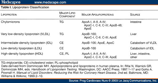
The apoproteins are located on the surface of the lipoprotein particles, while the lipids are in the centre. Phospholipids form the single layer shell of the particle, interspersed with apoproteins or free cholesterol. The lipoproteins can be broadly divided into two groups depending on the apoprotein they carry: those having ApoB, and those having ApoA. ApoB is also of two types: ApoB48 and ApoB100. While VLDL, LDL and IDL all carry ApoB100, CM carries ApoB48 which is an abridged form of ApoB100. ApoA is predominantly carried by HDL. Table 1 describes the details.
Relative particle size and structure of the lipoproteins is shown in Figure 5 and 6.
As in Table 1, each LP particle is associated with certain particular apoproteins. While Apo B- 48 is characteristic of CM, Apo B-100 is seen in VLDL and LDL. HDL is characterized by Apo AI and AII. Besides, Apo C and Apo E can shuttle between CM, VLDL and HDL. While Apo C is vital for functioning of LPL (lipoprotein lipase), Apo E is essential for uptake by liver through E-specific receptor recognition. LPL is the enzyme that breaks down TG into free fatty acids (FFA) and glycerol, thus allowing muscle cells and adipocytes to take up FFA from circulating CM and VLDL for energy and storage respectively.
While the function of CM, VLDL and LDL is to primarily transport lipids from the intestine and liver to the peripheral tissues, the HDL is primarily involved in reverse cholesterol transport. In this context, two other enzymes may also be mentioned here, LCAT and cholesterol ester transport protein (CETP). LCAT is present on surface of HDL and converts cholesterol into its esters, thus pushing the cholesterol on the surface into the interior of the particle. As HDL gathers more and more cholesterol from the blood vessel walls and from CM and VLDL, this enzyme helps the HDL transport the same by packing it into the inside or CETP is present in plasma and assists in transfer of TG present in VLDL and LDL with cholesterol esters present in HDL.9
Lipoprotein metabolism
There are two pathways of lipid metabolism, i.e., exogenous and endogenous.
Exogenous pathway
Dietary fats are broken down by the action of pancreatic lipase and bile juices, to form micelles and small fats particles which are taken up by the intestinal epithelium. After this uptake by the enterocytes, the micronized fat particles are packaged in conjunction with apolipoproteins, to form CM. These large lipoproteins contain apoprotein B48, and are taken up by the lacteals, being too large to be taken up by the vasculature. Passing through the chyle of the lymphatic system, the CM are carried into the systemic circulation by the Thoracic duct opening into the left Subclavian vein. In the blood, the CM gain apoproteins C2 and E from the HDL to become mature CM.
On coming into contact with the enzyme LPL located on the endothelial cells, the ApoC2 on the surface of the CM mediates the breakdown of the TG inside the CM into fatty acids and glycerol. The FA are taken up by the peripheral tissues and the glycerol is carried by the blood stream to the liver. Apo C2 is used up in the process, and the C2 deficient particle is now known as CM remnant. This particle then reaches the liver where it is recognized by the ApoE receptors on the surface of hepatocytes, and taken into the liver cells. Inside the hepatocytes, the particle is broken down into its component cholesterol esters, phospholipids (PL), vitamins A, D, E, K, as also amino acids (from the apoproteins). It may be noted that the ApoE on the surface of CM is important for the uptake of the CM remnant by the liver cells.
Endogenous pathway
This involves the synthesis, transport and utilization of VLDL, IDL, LDL and HDL. Let us describe VLDL metabolism first. VLDL, is characterized by the presence of Apoprotein B100. The VLDL particle is assembled in the liver and contains TG, CE, PL and vitamin E, along with ApoB100. After secretion from the liver, the VLDL particles gain Apo C2 and Apo E from the HDL, and reach all the tissues of the body, where the LPL recognizes the Apo C2 and initiates the breakdown of TG into FAs and glycerol. The VLDL loses the ApoC (which goes back to HDL) and is now known as IDL. This IDL particle can have two fates: (a) It can be taken up totally by the liver due to the presence of ApoE, (b) The hepatic lipase present on surface of liver cells can breakdown the triglycerides in the IDL remnant (during which process the ApoE will be used up), and the particle gets converted to LDL. The LDL particle thus contains only ApoB100.
REVERSE CHOLESTEROL TRANSPORT
While LDL is the main lipoprotein involved in carrying cholesterol from the liver to the peripheral tissues, HDL is responsible for the reverse process i.e., transporting excess cholesterol deposited primarily in the vessel walls back to the liver. While reverse cholesterol transport (RCT) is the major function of HDL, the latter also functions to prevent the oxidation of LDL which makes the LDL vastly more atherogenic. This antioxidant function of HDL is aided by the presence of the enzyme paraoxynase on its surface. Thus HDL is by and large an anti-atherogenic lipoprotein, though it contains significant amount of cholesterol.
The mechanism of RCT is as follows, the major apoprotein associated with HDL is Apoprotein A1 which is produced by intestinal cells and the liver hepatocytes. Apoprotein A1 is secreted into the blood in association with phospholipids and soon recruits enzymes LCAT, CETP and Paraoxonase. This particle also recruits many other apoproteins (but not ApoB100 and ApoB48) like ApoC and ApoE to form the particle known as discoidal nascent HDL.10
The discoidal nascent HDL travels to peripheral tissues and takes up cholesterol from tissues that have an excess of it. This is particularly so for the cholesterol that has accumulated in the macrophages in the arterial walls. The macrophages in the intima as also the foam cells give up their excess cholesterol by the action of ABCA1transporter (ATP binding cassette transporter), which transports the cholesterol to the nascent HDL. The nascent HDL has the enzyme LCAT on its surface which esterifies the cholesterol into cholesterol ester, which moves inside the HDL particle. Conversion of C to CE ensures that the cholesterol stays within the HDL right until it needs to be released, and the movement of CE into the center of HDL also changes the shape of nascent HDL from discoid to spherical. This CE rich HDL is known as HDL 3. The action of LCAT in the esterification process converts lecithin into lyso lecithin by the loss of a fatty acid.
The spherical HDL3 in the blood stream now exchanges its CE with the VLDL, IDL and LDL in return for triacylglycerol, courtesy the enzyme CETP. Thus the HDL gains TG and loses CE, and is now known as HDL2. This HDL2 has TG but continues to gain CE by the action of LCAT. HDL2 can have two fates: It can be taken up by hepatocytes in the liver through an apoA-1 mediated receptor interaction wherein the C and CE are separated and the modified HDL is again secreted into blood circulation to repeat the cycle. Alternately, the apoE in the HDL may bind to cell surface receptors on hepatocytes, resulting in the endocytosis of the lipoprotein particle, degradation of all constituents, and excretion of cholesterol into the intestinal lumen in the form of bile.
LIPOPROTEINS AND CANCER
Fast dividing cancer cells require cholesterol as an essential building block. Levels of LDL have been found to be raised in several cancers, including prostate, lung, ovaries and colon. HDL levels conversely, have been found to have a negative co-relation to prevalence of most cancers; this protective effect is presumed to be due to the anti-inflammatory properties of HDL. The intriguing apolipoprotein [a] has also been found to play a role in cancer progression; proteolytic breakdown products of this lipoprotein have been found to possess anti-angiogenic and anti-tumorogenic properties.11 Recently, lipoproteins have been studied for their potential role in targeted (chemotherapy) drug delivery in several cancers.
ALZHEIMER’S DISEASE
Alzheimer’s disease is characterized by deposition of Amyloid beta (Aβ) in the brain, can be affected by the association or otherwise of Aβ in the brain with lipidated apolipoproteins E and J.12 These latter are HDL like particles that influence the metabolic environment in the brain and CSF. Some studies indicate that functionally declined lipoprotein particles in the brain have an effect on the conversion of monomeric soluble Aβ 42 into oligomeric Aβ 42 peptides. Since clearance of the amyloid is hindered, there is deposition of these oligomeric fibrils in synapses and on neurons, affecting function.
LIPOPROTEINS AND THE LUNGS
ApoA-1 and HDL have been long known to have protective role in normal lung health, and also in a number of disease states like acute lung injury, COPD, pulmonary hypertension and viral pneumonia.13 Also, synthetic apoA-1 mimetic peptides have shown protective effects in animal experiments in models of asthma, pulmonary arterial hypertension and pneumonia. Thus, new treatments may be designed for lung disorders from apoA-1 mimetic peptides.
Another interesting observation that has surfaced is the positive association of pulmonary function with lipoprotein a [Lp(a)]. A recent study in the Free University of Berlin, by Nikolaus Buchmann revealed a positive correlation between lung volume and LDL-cholesterol in women.
LIPOPROTEINS AND IMMUNITY
Lipoproteins have been found to play a role in combating certain viral and bacterial pathogens, thus behaving as components of the innate immune system. This anti-infective role may be a result of direct attack on the microbe or by preventing the ingress of the pathogen into host cells. The best studied example in this respect is the role of HDLs in conferring immunity against Trypanosoma brucei in humans.14 Experimental evidence is accumulating that suggest high levels of circulating lipoproteins exert a protective role against respiratory and gastrointestinal infections.
Viral infections
HDL particles have been found to have non-specific and broad-based anti-viral activity against a wide spectrum of enveloped as well as non-enveloped DNA and RNA viruses.15 Examples include Poliovirus, Japanese Encephalitis Virus, Herpes simplex virus and Rubella virus.
Bacterial infections
Several classes of lipoproteins have the ability to neutralise lipopolysaccharides from gram negative bacteria.16 Experimental infusion of synthetic HDL particles has been shown to have a protective role against Gram negative bacteremia and endotoxic shock. These reconstituted lipoprotein particles (rHDL) have also been shown to blunt the LPS induced activation of the coagulation cascade. Lipoproteins have also been shown to inactivate lipotechoic acid and alpha-toxin from Staphylococcus aureus.
The anti-infective role of lipoproteins is postulated to be due to the following mechanisms:
- • Inhibiting the entry of intracellular pathogens into host cells, especially in the case of viruses, by interfering with viral attachment to host cells and subsequent penetration.
- • Inactivating bacterial toxins like LPS and enhancing their clearance. LPS binding with LDL causes fatty acyl chain in LPS to be incorporated into the phospholipid surface of LDL, thus masking the active sites of LPS.
- • Lysis of certain pathogens like Trypanosoma brucei, by targeting the parasites within the acidic parasitophorous vacuoles of macrophages that have ingested the parasite.17
- • Promoting opsonization, for example in Streptococci, by interaction of LDL with a scavenger receptor expressed in monocytes and Streptococcal protein 1. Opsonization then enhances phagocytosis.
- • Activation of complement system, for example in the killing of the gut pathogen Yersinia enterocolitica.
- • Chemical modification of lipoprotein, notably oxidative changes in LDL, brought about by ROS (reactive oxygen species). OxLDL then upregulates scavenger receptor expression in macrophages, which enhances phagocytosis of bacteria.
- • Redistribution of lipids to immune cells, by providing lipid substrates to macrophages and cholesterol for proliferation of lymphocytes, since cholesterol is required for new membrane synthesis.
SERUM LIPID INVESTIGATIONS
The whole purpose of the above discussion was to help us gain an insight into the actual significance of the values we see in a lipid profile report. From an era when cholesterol values were considered to be of overarching significance regarding the risk of cardio-vascular diseases, we have now reached a stage where we take into account the ratios between TG and cholesterol, and the total cholesterol to HDL ratios. There is also work going on to determine particle numbers, as for e.g. HDL and VLDL particle counts as opposed to total HDL or TG. The immediate future will see much technological sophistication in these assessments.
CONCLUSION
The significance of bodily lipids and the functions of lipoproteins are a subject of ongoing investigations. While cholesterol was much maligned in the past, recently the FDA has relaxed the restrictions on this vital lipid. Since the human brain is predominantly composed of lipids, it is of extreme importance to understand the significance of the dietary lipids. The long held theories of mechanical deposition of lipids in the vessel walls as the cause of cardio-vascular diseases have recently yielded to more plausible mechanisms linked to inflammation and plaque stability. Immune pathways are now believed to play a significant role in atherosclerosis, leading to much speculation on vaccine modalities to combat vascular disorders. Research into lipids and lipoproteins is thus a promising and exciting field, since cardio-vascular disorders are a major cause of morbidity and mortality in the current age.
Funding: No funding sources
Conflict of interest: None declared
Ethical approval: Not required
REFERENCES
- 1. Hussain MM. Intestinal Lipid Absorption and Lipoprotein Formation. Current Opinion in Lipidolgy. 2014;25(3):200-6.
- 2. Ball DW, Hill JW, Scott RJ. The Basics of General, Organic, and Biological Chemistry. 2011;1:17.2.
- 3. Worthington V, Cholesterol Esterase, Worthington Enzyme Manual, 1993. Available at: http://www.worthington-biochem.com:8080/resources/images/enzyme-manual/CEPM/reaction.jpg. Accessed on 1 September 2019.
- 4. Pimentel G, "Hydrogen bond", Wikipedia, The Free Encyclopedia. Available at: https://en.wikipedia.org/w/index.php?title=Hydrogen_bond&oldid=910405 940. Accessed on 1 September 2019.
- 5. Brian L. “Kansas State University Human Nutrition (FNDH 400) Flexbook”. NPP eBooks. 20018;19:199. Available at: https://newprairiepress.org/ebooks/19 . Accessed on 1 September 2019.
- 6. Kingsbury KJ, Bondy G, Understanding the Essentials of Blood Lipid Metabolism. Table I. Available at: https://img.medscapestatic.com/fullsize/migrated/451/762/pcn451762.tab1.gif . Accessed on 1 September 2019.
- 7. Zamora A, Lipoproteins Good cholesterol (HDL), Bad cholesterol (LDL). Available at: https://www.scientificpsychic.com/health/lipoproteins-LDL-HDL.html. Accessed on 1 September 2019.
- 8. Judström-Kareinen I, Mast Cells and HDL – Studies on Cholesterol Efflux and Reverse Cholesterol Transport - Scientific Figure on Research Gate. Available at: https://www.researchgate.net/figure/Schematic-drawing-of-lipoprotein-structure-Information-derived-from-Champe-et-al-2005_fig2_282356824. Accessed on 1 September 2019.
- 9. Siri-Tarino PW, Krauss RM. The early years of lipoprotein research: from discovery to clinical application. J Lipid Res. 2016;57(10):1771-7.
- 10. Chang TY, Yamauchi Y, Hasan MT, Chang C. Cellular cholesterol homeostasis and Alzheimer’s disease. J Lipid Res. 2017;58(12):2239-54.
- 11. Saba AB, Ajibade T, Role of Lipoproteins in Carcinogenesis and in Chemoprevention, Lipoproteins– Role in Health and Diseases. 2012;27:647-62.
- 12. Gordon EM, Figueroa DM, Barochia AV, Yao X, Levine SJ. High-density Lipoproteins and Apolipoprotein A-I: Potential New Players in the Prevention and Treatment of Lung Disease, Front Pharmacol. 2016;7:323.
- 13. Hajduk SL, Hager KM, Esko JD. Human high density lipoprotein killing of African trypanosomes. Annu Rev Microbiol. 1994;48:139-62.
- 14. Ravnskov U. High cholesterol may protect against infections and atherosclerosis. QJM. 2003;96:927-34.
- 15. Emancipator K, Csako G, Elm RJ. In-vitro inactivation of bacterial endotoxin by human lipoproteins and apolipoproteins. Infect Iminun. 1992;60:596-601.
- 16. Samanovic M, Molina-Portela MP, Chessler ADC, Burleigh BA, Raper J. Trypanosome lytic factor, an antimicrobial high-density lipoprotein, ameliorates Leishmania infection. Plos Pathog. 2009;5:e1000276.